

Painful biopsies, in which suspicious skin is lopped off and sent to a laboratory to detect skin cancer, may soon be a thing of the past. A study reveals a new technique that allows patients to have their skin examined under a special microscope, which can detect cancerous cells in just a few minutes.
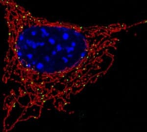
Caption/Image Credit: NICHD/U. Manor
The high-resolution device, called a multiphoton microscope, is able to see and identify a patient’s mitochondria, or the powerhouse of a cell. The study’s lead investigator, Irene Georgakoudi said that the mitochondria, “often form a beautiful network inside cells.”
When cancer is present in the body, it disrupts this network and makes the mitochondria unorganized, said Georgakoudi, also an associate professor of biomedical engineering at Tufts University in Medford, Massachusetts. When doctors observe mitochondria, they can diagnose skin cancer and other disorders based on what they see.
Multiphoton microscopes work by using photons, which are particles of light, allowing researchers to see the nicotinamide adenine (NADH) molecule. NADH is present in most cells, and it glows when placed under a light of a specific wavelength.
No Biopsy Dyes or Infection Risks
Currently, when doctors take biopsies of the skin to diagnose skin cancer, the sample is sent to a laboratory where doctors inject it with dyes and inspect it under a microscope for abnormal cells.
In some screening cases, the patients themselves are injected with dyes. But, because multiphoton microscope detects the cancerous cells through a special light, it bypasses this step.
“When NADH is in the mitochondria, it gives off a strong signal” that helps researchers detect it, Georgakoudi said. She added that because of NADH’s unique properties, researchers don’t have to inject the patient with dyes that would highlight the mitochondria.
Another downside of biopsies is the risk of infection or scarring to patients because of the cut that is made to extract the skin. Using imaging from a special microscope helps to avoid this situation.
How The Painless Technique Was Tested
Ten people with skin cancer, ranging from melanoma to the less invasive basal cell carcinoma, plus four with healthy skin, all had images taken of their skin with the microscope. Georgakoudi said data from 17 diseased locations and 12 healthy locations was observed.
The minute-long imaging session “didn’t cause any pain or discomfort to the patient,” Georgakoudi said. “We analyzed the images, in an automated way that requires only a couple of additional minutes, to characterize the manner in which mitochondria organize.”
The multiphoton microscope technique is not yet available to patients. However,if it works successfully in large studies, this new advancement in skin cancer detection technology could be available to the general population soon.
Ronke Idowu Reeves is a writer and journalist who hails from Brooklyn, NY. Her news and entertainment stories have appeared on WABC-TV-New York, Fox News Channel, VH1, BET.com plus in Sundance Film Festival’s Sundance Daily Insider and People Magazine.
![How To: ‘Fix’ Crepey Skin [Watch]](https://cdn.vitalupdates.com/wp-content/uploads/2017/05/bhmdad.png)


