

For parents who want to know more about what their unborn child does in the womb, a new technology could help that desire become a reality.
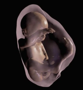
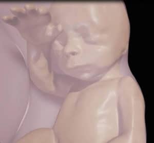
Research presented at the annual meeting of the Radiological Society of North America tells of the new technology that transforms MRI and ultrasound data into a 3D virtual reality model of a fetus. Study co-author Heron Werner Jr. from the Clínica de Diagnóstico por Imagem in Rio de Janeiro, Brazil said the technology could give new insight into how an unborn baby develops.
“The 3D fetal models combined with virtual reality immersive technologies may improve our understanding of fetal anatomical characteristics and can be used for educational purposes and as a method for parents to visualize their unborn baby,” he said in a press release.
Brazilian researchers created virtual reality 3D models based on fetal MRI results, then constructed the model using sequentially-mounted MRI slices. A physician begins the segmentation process by selecting body parts to be reconstructed in 3D.
The researchers have also been able to recreate a fetus’s entire internal structure as well, which could help doctors in assessing abnormalities. Werner and his colleagues used the Oculus Rift 2 headset, which places the user in an immersive environment. Users can hear the fetus’s heartbeat and study the 3D anatomy by just moving their head.
The 3D imaging has many potential applications, including assessment of whether the fetal airways are open or blocked. The development of fetal airways is an important issue and the 3D images could help physicians make better informed decisions about the delivery process.
“The physicians can have access to an immersive experience on the clinical case that they are working on, having the whole internal structure of the fetus in 3D in order to better visualize and share the morphological information,” Werner said. “We believe that these images will help facilitate a multidisciplinary discussion about some pathologies in addition to bringing a new experience for parents when following the development of their unborn child.”
Researchers have used the technology on patients at a clinic in Rio de Janeiro, including cases where a fetus had abnormalities present that required postnatal surgery.
After the original 3D ultrasound was created, it caused a wave of ultrasound “shops” to open, taking pictures for parents with no medical information, said Deborah Levine, professor of radiology at Beth Israel Deaconess Medical Center and Harvard Medical School.
“The Brazilian images are beautiful, but do you need those beautiful images to alter your treatment plan?” Levine said to NBC. “Those shops are still around, and they clearly go against prudent use of ultrasound in pregnancy, but people pay to have pictures taken of their fetuses.”
One concern is that both prolonged ultrasounds and MRIs can generate heat that can be harmful to the fetus. MRIs are also more expensive and more difficult to do than ultrasounds, usually reserving them for cases where there’s a real medical question.
The fetal MRIs cost at least $1,000 or more, and Levine said she doesn’t see the potential for 3D MRI parties.
“While the Brazilian model is technically feasible, it would not really be available to the average parent,” she said. “I’m not jumping up for joy on this one because I think we should use medical imaging only when it is medically needed.”
Werner said the 3D imaging goes beyond the average ultrasound.
“It provides fetal images that are sharper and clearer than ultrasound and MR images viewed on a traditional display,” he said to NBC.
Tori Linville is a freelance writer and editor from Clarksville, Tennessee. When she isn’t writing or teaching, she’s faithfully watching her alma mater, the University of Alabama, dominate the football field.
![How To: ‘Fix’ Crepey Skin [Watch]](https://cdn.vitalupdates.com/wp-content/uploads/2017/05/bhmdad.png)


